Fluorescence Detector
Bücker, R., Perrin, A., Manz, S., Betz, T., Koller, C., Plisson, T., Rottmann, J., Schumm, T. & Schmiedmayer, J.
Single-particle-sensitive imaging of freely propagating ultracold atoms
New Journal of Physics, 2009, Vol. 11(10), pp. 103039
We present a novel imaging system for ultracold quantum gases in expansion. After release from a confining potential, atoms fall through a sheet of resonant excitation laser light and the emitted fluorescence photons are imaged onto an amplified CCD camera using a high numerical aperture optical system. The imaging system reaches an extraordinary dynamic range, not attainable with conventional absorption imaging. We demonstrate single-atom detection for dilute atomic clouds with high efficiency where at the same time dense Bose–Einstein condensates can be imaged without saturation or distortion. The spatial resolution can reach the sampling limit as given by the 8 mum pixel size in object space. Pulsed operation of the detector allows for slice images, a first step toward a three-dimensional (3D) tomography of the measured object. The scheme can easily be implemented for any atomic species and all optical components are situated outside the vacuum system. As a first application we perform thermometry on rubidium Bose–Einstein condensates created on an atom chip.
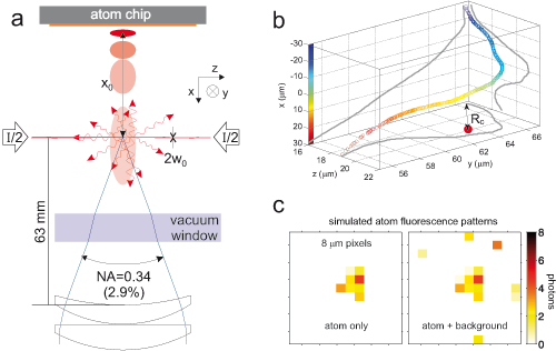
 (a) Schematic of the light sheet system. The sheet (waist in the x -direction w0 = 20 μm) is formed by two counterpropagating laser beams, each beam carrying half the total intensity I. After falling a distance x0 ∼10mm (not drawn to scale) the expanding atom cloud pierces the sheet and emits fluorescence photons. Outside the vacuum, an objective with numerical aperture (NA) of N = 0.34 collects 2.9% of the light and transmits it to a camera. (b) Typical trajectory of a single atom falling through the sheet, obtained by Monte Carlo simulation. The atom performs a random walk in momentum space due to the stochastic photon absorption and emission events. This random walk translates into real space on a typical scale of 10 μm. The colour encodes the vertical position of the atom. In the horizontal plane, the red dot indicates the transverse centroid position of the emitted photons, which is located at a distance Rc from the initial atom position. (c) Simulated image of a typical single atom, including effects of diffusion, signal amplification and lens aberration (left) and background (right).
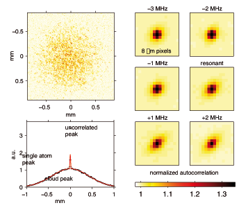
 Image autocorrelation and single-atom molasses effect. (a) Typical image of a thermal cloud (fully integrated along the vertical direction) using the light sheet system. (b) Cut through the y-axis of the 2D autocorrelation function of (a). Three features can be distinguished which correspond to photon shot noise, single atoms and the cloud envelope. A double Gaussian fit is shown. (c) Central peak of the normalized 2D autocorrelation function for thermal cloud images at different detuning settings of the light sheet. The scattering rate per atom was held constant by adjusting the intensity in the sheet. The anisotropy of the peak depends on the detuning, indicating a 1D Doppler cooling during the imaging process. Arrows indicate the direction of the light sheet (LS).
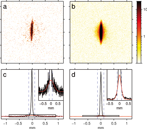
 (a) Single-shot image of a 400μs time-slice at the centre of a quasi-pure Bose–Einstein condensate. (b) Averaged image of a quasi-pure Bose–Einstein condensate over 20 realizations. (c) Longitudinal profile of (a). Even though the thermal fraction is very small, its shape can still be fitted in a single experiment run due to the high dynamic range of the fluorescence detector. We deduce a temperature of 220 (150) nK and estimate the thermal fraction to include the fluorescence scattering of about 100 atoms. The inset shows a zoom into the shape of the thermal fraction. (d) Longitudinal profile of (b). By fitting the thermal fraction we are able to obtain a temperature of only 64 (15) nK. In panels (c) and (d), the vertical dashed lines indicate the boundary of the excluded region of the fit.